
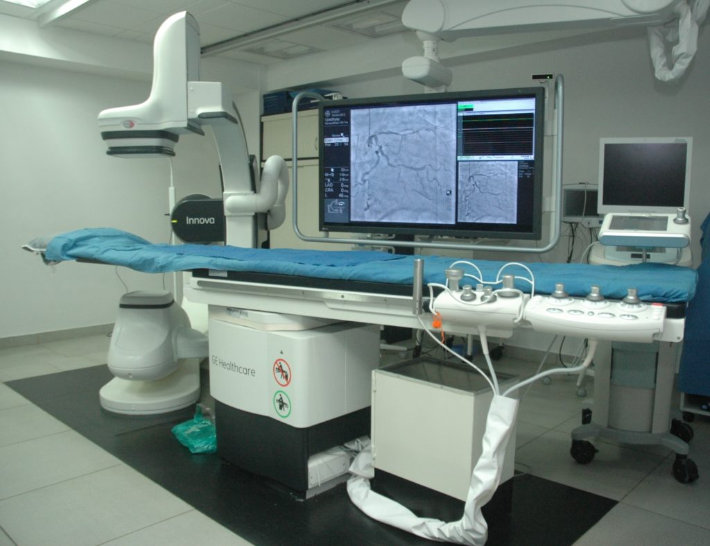
Coronary angiogram helps the doctor determine the extent and the seriousness of the atherosclerosis in the arteries in the heart and determine the mode of treatment to be carried out. It also offers detailed images of the blood vessels in the heart and this is especially useful when a surgical operation is being thought about.
Coronary Angioplasty A coronary angioplasty is performed using local anaesthetic, when the catheter is in place, a thin wire is guided down the length of the affected coronary artery, delivering a small balloon to the affected section of artery. This is then inflated to widen the artery, squashing fatty deposits against the artery wall so blood can flow through it more freely when the deflated balloon is removed. If a stent is being used, this will be around the balloon before it's inserted. The stent will expand when the balloon is inflated and remains in place when the balloon is deflated and removed. A coronary angioplasty usually takes between 30 minutes In most cases, the blood flow through the coronary arteries improves after an angioplasty. Many people find their symptoms get significantly better and they're able to do more than they could before the procedure. If you've had a heart attack, an angioplasty can increase your chances of surviving more than clot-busting medication (thrombolysis). The procedure can also reduce your chances of having another heart attack in the future.
Renal angiogram helps the doctor determine the extent and the seriousness of the atherosclerosis in the arteries in the renal and determine the mode of treatment to be carried out. It also offers detailed images of the blood vessels in the kidney and this is especially useful when a surgical operation is being thought about.
Renal angioplasty A Renal angioplasty is performed using local anaesthetic, when the catheter is in place, a thin wire is guided down the length of the affected renal artery, delivering a small balloon to the affected section of artery. This is then inflated to widen the artery, squashing fatty deposits against the artery wall so blood can flow through it more freely when the deflated balloon is removed. If a stent is being used, this will be around the balloon before it's inserted. The stent will expand when the balloon is inflated and remains in place when the balloon is deflated and removed. A renal angioplasty usually takes between 30 minutes In most cases, the blood flow through the Renal artery improves after an angioplasty. Many people find their symptoms get significantly better and they’re able to do more than they could before the procedure.
Peripheral angiogram helps the doctor determine the extent and the seriousness of the atherosclerosis in the arteries in the periphery and determine the mode of treatment to be carried out.
Peripheral angioplasty is performed using local anaesthetic, when the catheter is in place, a thin wire is guided down the length of the affected Peripheral artery, delivering a small balloon to the affected section of artery. This is then inflated to widen the artery, squashing fatty deposits against the artery wall so blood can flow through it more freely when the deflated balloon is removed. If a stent is being used, this will be around the balloon before it's inserted. The stent will expand when the balloon is inflated and remains in place when the balloon is deflated and removed. A Peripheral angioplasty usually takes between 30 minutes In most cases, the blood flow through the Peripheral artery improves after an angioplasty In most cases, the blood flow through the peripheral arteries improves after an angioplasty. Many people find their symptoms get significantly better and they’re able to do more than they could before the procedure. If you've had a claudication, an angioplasty can increase your chances of relieving symptoms. The procedure can also reduce your chances of having symptoms of PVD in the future.
Four vessel angiogram helps the doctor determine the extent and the seriousness of the atherosclerosis in the arteries in the brain & neck and determine the mode of treatment to be carried out. It also offers detailed images of the blood vessels in the brain & neck and this is especially useful when a surgical operation is being thought about.
Carotid /vertebral angioplasty angioplasty is performed using local anaesthetic, when the catheter is in place, a thin wire is guided down the length of the affected Carotid /vertebral artery, delivering a small balloon to the affected section of artery. This is then inflated to widen the artery, squashing fatty deposits against the artery wall so blood can flow through it more freely when the deflated balloon is removed. If a stent is being used, this will be around the balloon before it's inserted. The stent will expand when the balloon is inflated and remains in place when the balloon is deflated and removed. A 5. Carotid /vertebral angioplasty usually takes between 30 minutes In most cases, the blood flow through the Carotid /vertebral artery improves after an angioplasty Many people find their symptoms get significantly better. If you've had a stroke, an angioplasty can increase your chances of relieving symptoms. The procedure can also reduce your chances of having another stroke in the future.
Device Closure is a minimally invasive procedure that doesn’t require opening the chest, which means less pain and a quicker recovery time compared to traditional open-heart surgery.
Device Closure procedures are effective in repairing certain types of Congenital Heart Defects, such as Atrial Septal Defects (ASDs), Ventricular Septal Defects (VSD), and Patent Ductus Arteriosus (PDA). These defects can lead to symptoms such as shortness of breath, fatigue, and arrhythmia, which can be improved with the closure of the hole.
Since Device Closure procedures are minimally invasive, there is a lower risk of complications compared to Open-Heart Surgery.
The heart has 3 valves namely mitral aortic and tricuspid.Stenosis is a blockage of the valve in the heart in which the flaps of the valve (leaflets) become stuck together. This reduces the blood flow from one heart chamber to another, causing a back-up of fluid into the lungs. This makes you feel short of breath (puffed).A valvuloplasty is a procedure where the valve is widened using a balloon. This will allow the blood to flow more easily and relieves you from symptoms.
Endovascular Aneurysm Repair (EVAR) is a minimally invasive procedure used to treat abdominal aortic aneurysms (AAA)—a condition where the main artery in the abdomen weakens and bulges. Instead of open surgery, EVAR involves placing a stent graft through small incisions in the groin to reinforce the weakened artery and prevent rupture. This advanced technique leads to faster recovery, reduced hospital stay, and lower complication risks, making it a preferred treatment for eligible patients.
is a procedure for treating patients with valvar stenosis who are not considered candidates for standard open heart surgery. In this procedure, a new biological valve is inserted via a catheter (a thin tube) into the heart. The valve is inserted into your heart from your femoral artery/Vein, the main blood vessel in your groin. Patient gets Freedom from breathlessness, giddiness, weakness, increase in life expectancy, reduction in heart failure related hospitalisation.
Left Atrial Appendage Occlusion (LAAO) is a minimally invasive heart procedure designed to reduce the risk of stroke in patients with atrial fibrillation (AFib) who cannot take long-term blood thinners. The procedure involves sealing off the left atrial appendage (LAA), where blood clots often form in AFib patients. LAAO provides a safe, effective alternative to anticoagulant therapy, lowering stroke risk while improving quality of life.
Electrophysiology studies are performed to determine an arrhythmia diagnosis or the mechanism of a diagnosed arrhythmia the doctors can then suggest what is best for you. This may be insertion of a pacemaker or implantable cardiac defibrillator (ICD)or Radiofrequency ablation (RFA) or Medical management
A pacemaker is a device, you have had implanted under your skin to prevent heart beating too slow.
CRT D makes your heart chambers pump synchronously (at the same time). You may feel less breathless and have more energy. The main benefit of the defibrillator function is that it works to protect you from the risk of sudden cardiac death due to dangerously fast heart rhythms (VT or VF). The defibrillator can also treat some rhythm disturbances without you being aware of it by fast pacing, and without shocks.
AICD will help slow down a fast heartbeat. Sometimes the heart beats too fast. This does not allow the chambers of the heart to fill properly. If enough blood is not pumped around the body and it is untreated, it may lead to dizziness, fainting or loss of consciousness. This is a potentially life threatening condition. The AICD stops the fast heartbeat. This is done by ‘pacing’ the heart rapidly or by giving an electrical ‘shock’ to the heart.
An Inferior Vena Cava (IVC) filter is a small, metal device placed in the largest vein (IVC) in the body to prevent life-threatening pulmonary embolism (PE) by catching blood clots before they reach the lungs. This procedure is recommended for patients at high risk of clots who cannot take blood thinners. IVC filters provide immediate protection and can be either permanent or retrievable, depending on individual patient needs.
Sometimes the fatty substance that builds up in arteries (plaque) contains calcium that makes the blockage hard. If a plaque is severely calcified, a standard angioplasty balloon may not be able to cross the blockage and push it to the sides of the artery.Rotational atherectomy is a procedure that can be performed to drill through tough blockages. A tiny rotating cutting device is used to open a narrowed artery and improve blood flow. The pieces of plaque dislodged by the rotational atherectomy device are small enough to be absorbed by the blood stream.
FFR is a test used to measure how much blood flow is being restricted by a blockage in an artery. A special, pressure-sensing guidewire is fed through a catheter to the site of the blockage in the artery.
When water flows through a garden hose the flow is driven by the pressure in the tap. If there hose has no obstruction, the pressure at the end of the hose is the same as the pressure at the tap. In a healthy heart artery, the pressure at the end of the artery is the same as at the beginning of the artery (where it comes off of the aorta). But when a blockage reduces flow through the heart artery, or a garden hose, the pressure at the end is reduced proportionate to the restriction. The greater the restriction, the lower the pressure downstream because the flow is reduced. When the FFR wire is placed across the blockage, it measures the pressure in front of and beyond the blockage. If the blood pressure in the artery or beyond the blockage is found to be significantly reduced, then that blockage may be a good candidate for angioplasty and stenting to clear the blockage and prop the artery open.
All ultrasound tests, including IVUS, use sound waves to create images. IVUS is used to gather images of the inside of arteries to find out if a blockage is present, and if so, how serious the blockage is.
During an IVUS test, a catheter with an ultrasound probe at the end is threaded over a guidewire in the artery to the area to be tested. The ultrasound catheter sends out sound waves and receives echoes from the sound waves as they bounce back from the body’s tissues. These echoes are translated by a computer into images of the artery. IVUS is a test that may be performed during an angiogram (also known as cardiac catheterization). Your doctor may use IVUS if the blockages seen with the angiogram appear to be borderline-severe, or if the doctor needs more information about the plaque anatomy.
IVUS is useful because it allows the interventional cardiologist to measure the amount of plaque inside vessels as well as how much much space is available in the artery for blood to flow through. If it is determined that you need angioplasty to treat the blocked artery, IVUS can help with accurate positioning of the balloon and stent.
OCT is an imaging tool used to take high-resolution pictures of blood vessel walls. OCT provides interventional cardiologists with detailed images of plaque (cholesterol and other materials that have accumulated in the walls of the artery and can rupture, causing a blood clot to form at the site and block off critical blood flow). Like IVUS, this detailed information about plaque build-up in arteries can help interventional cardiologists determine where best to place stent
Shockwave intravascular lithotripsy (IVL) is an innovative medical technique adapted from the established treatment for kidney and ureteral stones. It involves the use of a percutaneous device to generate acoustic pressure waves, which are directed towards calcified lesions in blood vessels. These waves effectively break down both superficial and deep calcium deposits within the vessel walls. This process assists in preparing the artery for the successful deployment of a vascular stent, ultimately restoring proper blood flow and improving patient
Orbital Atherectomy (OA) is an innovative, minimally invasive procedure designed to treat severely calcified coronary arteries. It uses a high-speed, diamond-coated rotating burr to break down hardened plaque, allowing for improved blood flow and safer stent placement. OA is particularly beneficial for patients with complex coronary artery disease, where traditional balloon angioplasty alone may not be sufficient. This technique enhances procedural success, reduces complications, and improves long-term heart function, making it a crucial advancement in interventional cardiology. Patients undergoing OA typically experience faster recovery and better treatment outcomes.
Electrocardiogram (ECG) is a quick, non-invasive test that records the heart’s electrical activity. It helps in diagnosing conditions such as arrhythmias, heart attacks, and other cardiac abnormalities. This simple yet essential test is used for routine heart check-ups, detecting early warning signs, and monitoring heart health over time. ECG provides critical insights that aid in timely treatment, reducing the risk of serious complications.
2D Echocardiography (2D Echo) with Colour Doppler is a highly advanced ultrasound imaging technique that assesses the heart’s structure, function, and blood flow dynamics. It is particularly useful in detecting valvular heart disease, congenital defects, and cardiomyopathy. The Colour Doppler feature enhances visualization of blood flow abnormalities, helping doctors diagnose conditions with precision. This non-invasive, painless test plays a crucial role in early detection, treatment planning, and ongoing monitoring of heart health.
Dobutamine Stress echocardiography is a special type of Cardiac Ultrasound test to assess how well the heart works under stress. During test contractile &/or valvular function of the heart is assured.A Drug named Dobutamine is given intravenously according to the patient’s weight at incremental doses for pharmacological stress. Patient needs to be kept in hospital for 2-3 hrs. for cardiac monitoring. Intravenous line is taken to give medication during test
TEE is a special type of echocardiography of Heart. Pictures of the heart are taken from inside the body. This procedure gives better quality pictures of the heart. The equipment that takes the pictures is called the echocardiography probe. The back of your throat will be sprayed with a local anaesthetic which will make it easier to swallow the echocardiography probe. The probe is put into the mouth and it passes down to the oesophagus. The probe will be in place for about15 minutes until the test is completed. . The doctor will see the back of the heart from this position.At the end of the test, the probe will be removed.
If you can walk easily, you can walk on the treadmill. The speed and slope of the treadmill will increase every three minutes. This makes your heart do more work.. Your pulse, blood pressure and electrocardiogram are monitored during and after the test. It measures the function of the heart, lungs and blood vessels. It is done to help diagnose blocked arteries in the heart (coronary artery disease), assess abnormal heart beats or to check the function of pacemakers.
24-Hour Holter Monitoring is a continuous ECG recording that tracks heart rhythms over an extended period, typically 24 to 48 hours. It is essential for detecting irregular heartbeats, unexplained palpitations, dizziness, and silent arrhythmias that may not appear during a routine ECG. The portable device records every heartbeat during daily activities and sleep, providing detailed data for accurate diagnosis and treatment. This test is crucial in identifying hidden heart conditions, guiding treatment adjustments, and improving overall cardiac care.
24-Hour Blood Pressure (BP) Monitoring is an advanced diagnostic test that measures blood pressure at regular intervals throughout the day and night. It is especially useful in diagnosing hypertension, white coat syndrome, and masked hypertension. Unlike a single clinic measurement, this test provides a comprehensive picture of BP fluctuations, helping doctors assess medication effectiveness and lifestyle impact. By offering a more accurate blood pressure profile, this test aids in better diagnosis, personalized treatment, and long-term cardiovascular health management.